
A medical software for the electrophysiologist to extract key information from cardiac MRI & CT images
Plan the EP intervention in a fast and easy way with a rich variety of information obtained from cardiac imaging. While planning, you will learn from your patient data to decide the best treatment strategy.
Planning, intervention guidance or research. All can be done with ADAS 3D.
Decision making before intervention
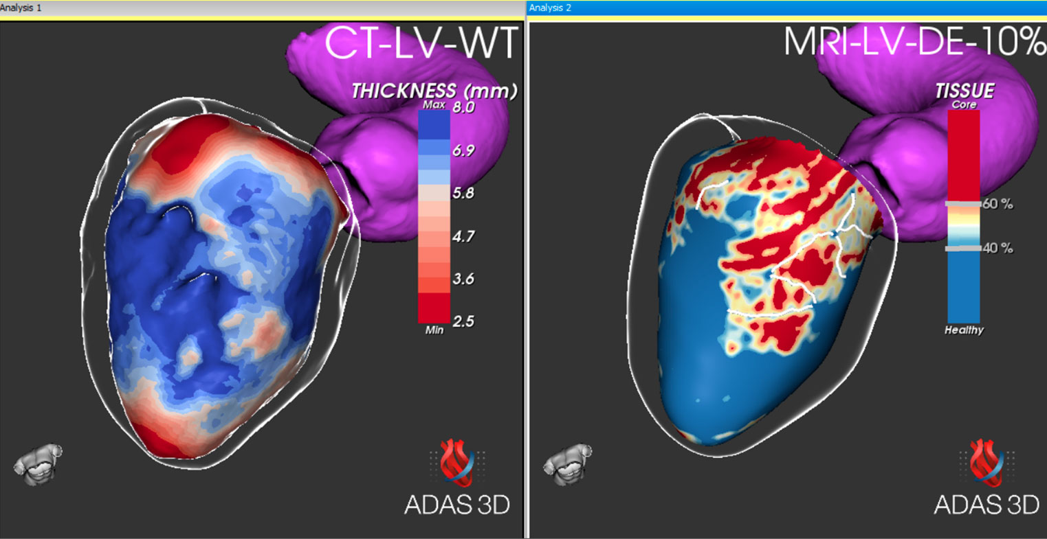
Understand the fibrosis distribution in the myocardium from MRI and the wall thickness and surrounding anatomical structures from CT.
During the procedure
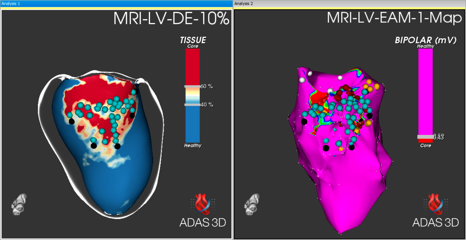
Use the ADAS 3D model as a roadmap to the catheter navigation system to accurately treat your patient.
Post-procedure revision
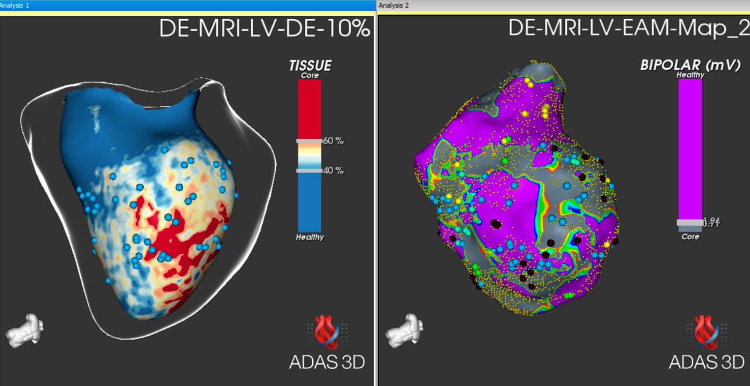
After the intervention, study the electroanatomical map with ADAS 3D and compare it with the MRI information to quantify outcomes.
How can ADAS 3D LV help you?
Help in deciding the approach
We help you to obtain additional information about your patient to decide the approach before the intervention.
Understanding of LV substrate
We help you to understand the LV substrate by visualizing and quantifying its fibrotic tissue distribution.
Increase the operator’s confidence
We help increase the operator’s confidence during navigation by providing a roadmap to assist in the procedure.
Obtain all the benefits from advanced imaging
We help you to connect to advanced imaging radiological services to import DICOM images for MRI.
Border-zone corridors automatically detection
Automatic transmural detection of border-zone corridors and the quantitative analysis for each corridor.
Easy-to-understand ‘layers’
Visualize the Left Ventricle in concentric layers going from endo to epicardium to appreciate the scar transmurality.
Tissue characterization
Obtain a 3D Cardiac Model Segmentation with an automatic classification of tissue using selectable imaging thresholds
Export to EP navigation system
Help during the intervention by exporting structures (including anatomy, fibrosis information and corridors) to formats compatible with EP navigation systems
How can ADAS 3D LA help you?
Help in identifying the gaps
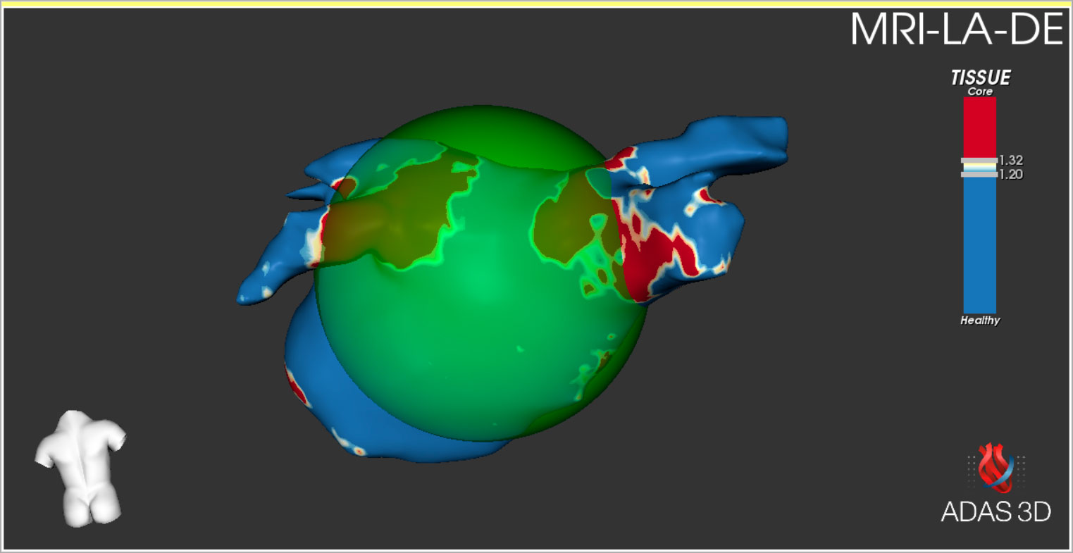
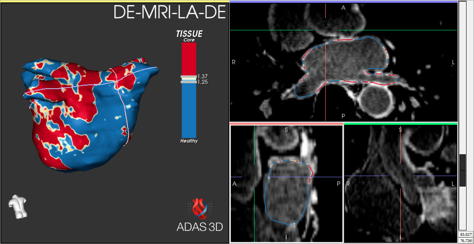
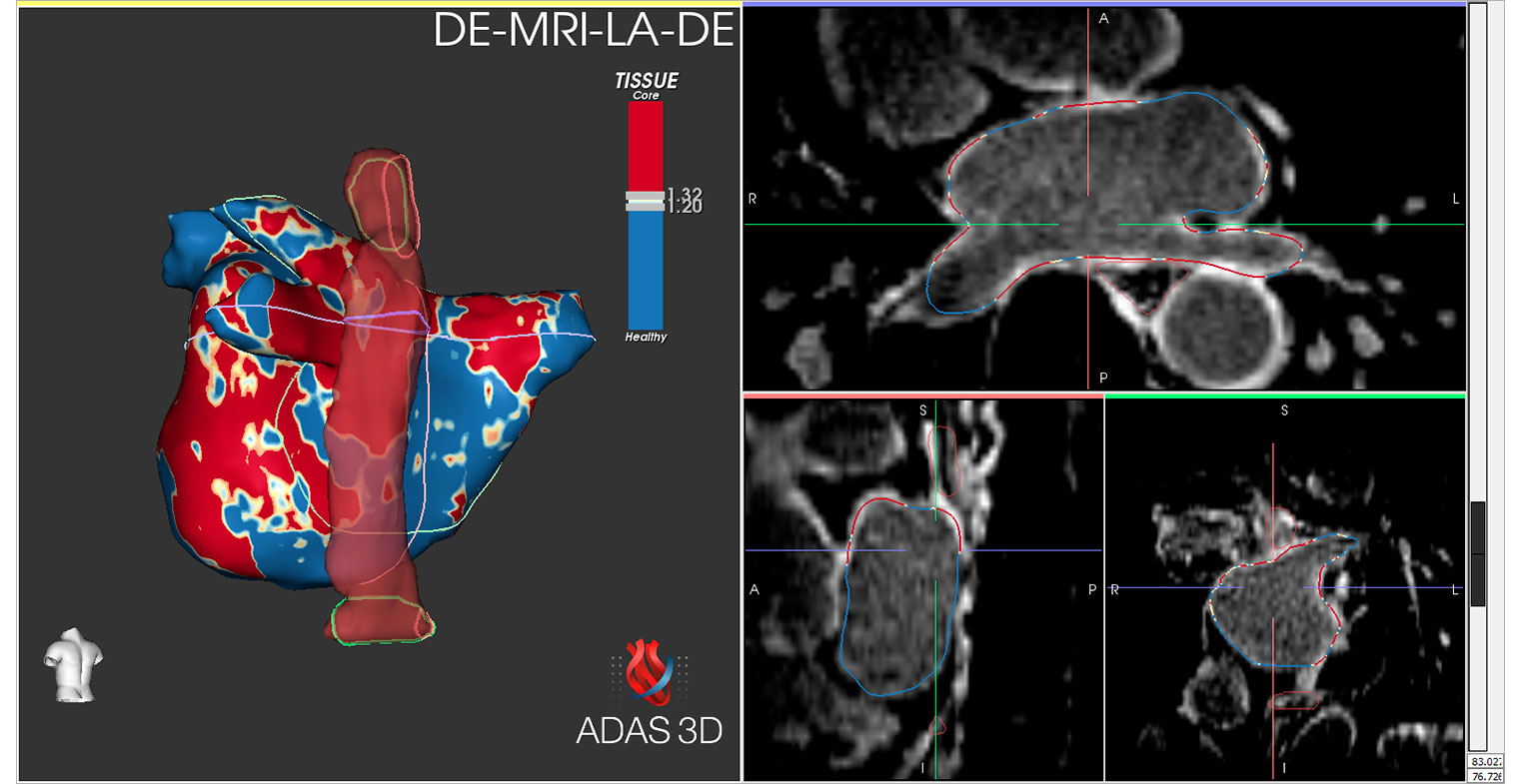
We help you differentiate the healthy tissue from the fibrosis, including the identification of gaps previously generated.
Understanding of fibrotic substrate
We help you to understand the LA substrate by visualizing and quantifying its fibrotic tissue distribution. Use this information to decide the approach before the intervention.
Increase the operator’s confidence
We help increase the operator’s confidence during navigation by providing a roadmap to assist in the procedure.
Obtain all the benefits from advanced imaging
We help you to connect to advanced imaging radiological services to import DICOM images for MRI.
Anatomical information
Visualize a 3D view of the left atrium and the surrounding anatomical structures.
Tissue characterisation
Obtain a 3D Cardiac Model Segmentation with an automatic classification of tissue based on Image Intensity Ratio using selectable imaging thresholds.
Export to EP navigation system
Help during the intervention by exporting structures (including anatomy and fibrosis information) to formats compatible with EP navigation systems
Analysis quantification
Quantify the amount of enhanced fibrosis in the LA to differentiate the fibrotic substrate from the healthy tissue.
Technical Specifications
Windows-based
The ADAS 3D software can be installed on a laptop or desktop computer (see minimum requirements).
Connection to PACS system
Download DICOM images and save cases from a central archive. This feature is available in the integrated version with Cvi42.
Floating licenses
ADAS 3D can be used on any computer within the same hospital under a single license. This feature is available in the integrated version with Cvi42.
Study database
Store and manage processed studies.

Pie Medical Imaging to lider w projektowaniu innowacyjnego oprogramowania wspierającego pracę lekarzy. Firma oferuje pakiety oprogramowania wspomagającego bezpieczne i skuteczne wykonywanie procedur z zakresu m.in. kardiologii, kardiochirurgii, chirurgii naczyniowej i radiologii.

MicroPort CardioFlow to firma produkująca sprzęt medyczny, zajmującą się badaniami, rozwojem oraz komercjalizacją innowacyjnych rozwiązań przezcewnikowego i chirurgicznego leczenia wad zastawkowych serca.

Narzędzie AI stworzone do wsparcia decyzji klinicznych.
Obszar Neuronaczyniowy, Kardiologiczny i naczyniowy, Radiologia, Edge Cloud oraz Life science
Pomoc lekarzom to nasza misja
Dane firmy:
mTree Medical Solutions Sp. z o.o.
Tychy, ul. Harcerska 60
NIP: 6462941906
REGON: 364059267
Dane kontaktowe:

